
Equine Ultrasound Pregnancy Images
All of the mare ultrasound pregnancy images and videos below were produced using a Universal S8 Color Doppler Ultrasound from Universal Imaging, Bedford Hills, NY.
Click on all images to see an enlargement
For videos you can go “full screen” by clicking the screen icon in the bottom right corner of each video. ![]()
Jump to day number:
11 12 13 14 15 16 17 18 19 20 21 22 23 24 26 27 33 40 43 53 58
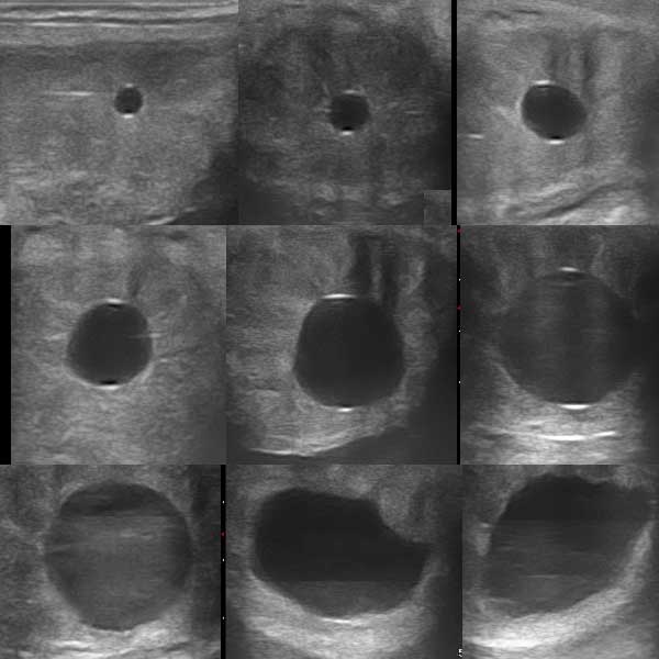
 Growth of the embryonic vesicle between days 10-17 is usually linear, averaging approximately 3.4 mm/day. In order to maintain a constant, the images below between days 10-18 are all of the same mare’s pregnancy. There is a slight “bump” in the linearity on day 14, but the overall average is maintained. A temporary plateau of growth and final cessation of mobility within the uterus commences around day 17. This plateau of cross-sectional conceptus diameter, showing only a marginal increase of diameter, if any at all, will continue until around day 28 by which time the yolk sac (of which the early vesicle primarily consists) is regressing in size, while the allantoic sac is increasing in diameter. Although the uterine horn cross-sectional diameter remains constant, the conceptus is in fact undergoing longitudinal elongation up the uterine horn, which can be visualised by placing the transducer in a longitudinally along the uterine horn.
Note that on each of the images from day 10-15 there appears at both the top and the bottom of the conceptus a small white area. This is thought by some to be a definitive indication that this is a pregnancy rather than a cyst or other fluid-filled area of the uterus, but that is incorrect. The small white area doesn’t even physically exist! It is called a spectral reflection and is an artefact of ultrasonography which may be seen on any enclosed area of fluid being ultrasounded.
Mare Ultrasound Pregnancy Images


Around day 17 – often exactly to the day – a loss of regularity of shape of the vesicle occurs in conjunction with cessation of movement through the uterus. This is primarily as a result of the conceptus increasing in diameter to a point greater than the internal diameter of the uterine horns. This cessation of movement is often erroneously referred to as “implantation”, but there is no invasion of the endometrium (the uterine lining) at this stage, and the correct term is “fixation”. The loss of regularity of shape has been seen to be a cause of concern by some – even to the point of belief of impending pregnancy loss and treatment with prostaglandin to “start again” – but it is absolutely normal at this stage. Concern is sometimes compounded by the belief that the mare is a few days earlier post-ovulation than she truly is, the duration calculation having been made from the time she was last positively responsive to the stallion. Following estrus, mares do not become resistant to a stallion until progesterone levels are adequately elevated to cause that resistance, and the progesterone source is the developing Corpus Luteum (“CL”) which forms following ovulation. Resistance to the stallion may therefore not occur until as much as 48 hours after ovulation, although up to 24 hours is more common. When viewing mare ultrasound pregnancy images calculated as being 15 days from that point could therefore mean she is in reality as much as 17 days post-ovulation and conception – and in the time frame when loss of regularity of shape is to be expected.
Day 17

17 day image

17 day measured


Normally, around day 20-21, when using standard ultrasound equipment, the “embryo proper”, as it is termed, will become visible as a small irregularity on the border of the vesicle – most commonly towards the ventral (bottom) region. With this particular ultrasound unit, which has images of superior quality, we can just see the embryo proper in the 19 day image above as a small whiter speck on the ventral region of the conceptus (arrowed in red below). By 20 day with this unit, it is very clearly visible in the image, albeit on the lower right margin of the vesicle rather than the more normal ventral region.

Embryo proper (arrow)
By day 22-23, again when using standard ultrasound equipment, the heartbeat should become visible, showing as a tiny flicker of grey-scale change. When using Color Doppler ultrasound, while the embryo itself may not be visible much earlier, the heartbeat can often be seen sooner, as evidenced by the 20 day CD video below which shows the brief flicker of color of the heartbeat.
The use of Color Doppler in the ultrasound indicates movement within the view. With the dual-color mode, two colors are seen – blue and orange – one indicating movement towards, the other movement away from the probe. What one is seeking is the movement of blood – blood flow. If however the mare herself moves, or the probe is moved within the mare, there is an artificial suggestion of movement evidenced by large areas of blue or orange. When seeking a heartbeat in an embryo, it is therefore imperative that the mare and the probe remain still. In the still CD 20-day image below, one can clearly see an orange speck within the embryo, which is indicative of a heartbeat. It is also visible as one quick orange flash at the beginning of the 20-day CD video. The embryonic heartbeat can be differentiated from the maternal blood flow in the surrounding vessels, as the former is significantly faster.


By day 26, the newly emerging allantoic sac can often be seen below the embryo proper – a process started somewhere around 22-24 days. This will increase in size, while the yolk sac (which is above the embryo) will decrease in size resulting in an apparent “floating upwards” of the embryo within the vesicle. It is worth comparing this 26 day image with the 27 day image immediately below it, where one can see a dramatic difference in the size of the allantoic sac. This difference may be as a result of different rates of progression in different mares, as well as transducer angle. It is worth while remembering that while there are averages, there are no absolute standards!


27 day image

27 day measured
The comparison in size of the allantoic sac (lower) and yolk sac (upper) is clearly visible, with the former increasing in size causing the embryo to appear to “rise” in position.
Day 27 (second mare – longitudinal view)

27 day image

27 day CD image

27 day measured
27 day CD video


The allantoic sac has now enlarged and elevated the embryo dorsally (upwards) while the yolk sac has almost completely regressed. By around 36 days, the embryonic ascent will be complete, and the umbilical cord will be forming at the dorsal pole. Blood flow within the embryo is clearly visible in the color doppler image. Around this time, the endometrial cups will have formed in the region of the chorionic girdle which forms where the allantoic and yolk sacs meet. The endometrial cups are responsible for the secretion of the hormone equine chorionic gonadotropin (eCG) which in turn is responsible for promotion of the production of secondary CLs which will increase progesterone to assist pregnancy maintenance until around 100 days of pregnancy. At 40 days the term for the developing foal will change from “embryo” to “fetus” in recognition in part of the changed state of nutritional source as the umbilicus is formed, and the allantochorion commences attachment to the endometrium, becoming the placenta – a process which is not completed until around 140 days of pregnancy. In the near future the size of the fetus will preclude its being entirely visible in mare ultrasound pregnancy images.


The internal bloodflow of the fetus is now clearly visible, as well as some umbilical flow. The small remnant of the yolk sac can be seen still attached to the left.

43 day image

43 day CD image

43 day measured
The internal blood-flow of the fetus is clearly visible, as well as umbilical flow. The small remnant of the yolk sac can be seen still attached to the left.

53 day fetal side view

53 day area measured

53 day length measured
This side view of the fetus clearly shows the head at the top, with limbs being visible.

58 day image

58 day CD image

58 day measured
58 day CD video. The fetal heartbeat and umbilical blood flow are clearly visible.
© 2014, Equine-Reproduction.com, LLC
Use of article permitted only upon receipt of required permission and with necessary accreditation.
Please contact us for further details of article use requirements. Other conditions may apply.

